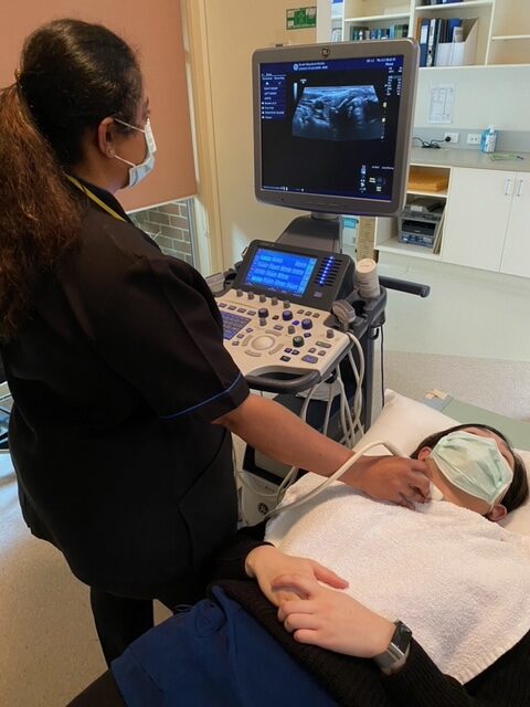
Ultrasound

About Ultrasound

Ultrasound (US) imaging, also called ultrasound scanning or sonography, is a painless, fast, and easy method of “seeing” inside the human body with high-frequency sound waves. The sound waves are recorded and displayed as a real-time visual image. No ionizing radiation is involved in ultrasound scanning.

In most ultrasound examinations, a transducer, a lightweight device that produces sound waves is placed on the patient’s skin. There are also special transducers, which can be put into the vagina or rectum to image these areas of the body.

Common Uses

  • Abdominal organs(liver, gallbladder, spleen, pancreas, kidneys, bladder)
  • Pelvic organs(prostate, uterus and ovaries)
  • During pregnancyto monitor the development of the embryo or foetus.
  • Superficial organs(breast, thyroid, joints)
  • Heart
  • Blood Flow.Doppler ultrasound is a special technique used to examine blood flow. Doppler images can help to see and evaluate blockages to blood flow, such as clots, and build-up of plaque inside the vessels.
  • Biopsy.It can also be used to guide procedures such as needle biopsies, in which a needle is used to sample cells from an organ for laboratory testing.

Preparation

  • You should wear comfortable, loose-fitting clothes.
  • For gallbladder examination, a fasting period of 4-6 hours is required to visualize your gallbladder, otherwise no fasting is required.
  • For pelvic ultrasound, you may be asked to drink up to four glasses of water prior to your exam and avoid urinating, so your bladder is full when the scan begins.
  • Please be punctual for your appointment and please bring your referral form Medicare card / concession card.

Procedure

  • You will lie on your back on an examination table.
  • A clear gel is applied to your body in the area to be examined, to help the transducer make secure contact with the skin. The sound waves produced by the transducer cannot penetrate air, so the gel helps eliminate air pockets between the transducer and the skin. The gel is water soluble, safe and harmless and can easily be wiped off after the scan with a paper towel.
  • The sonographer presses the transducer firmly against the skin and sweeps it back and forth to image the area of interest.
  • For pelvic ultrasound, it may be done transabdominal or transvaginal to see the uterus and ovaries.
  • When the examination is complete, the gel can be easily cleaned with the use of tissue.

Benefits and Risks

BENEFITS

  • Ultrasound scanning is non-invasive (no needles or injections in most cases) and is usually painless.
  • Ultrasound is widely available and easy to use.
  • Ultrasound imaging uses no ionising radiation and is the preferred image modality for diagnosis and monitoring of pregnant women and their unborn infants.
  • Ultrasound provides real-time imaging, making it a good tool for guiding minimally invasive procedures such as needle biopsies.
  • Ultrasound images can visualise structures, movement and live function in the body’s organs and blood vessels.

RISKS

  • For standard diagnostic ultrasound, there are no known harmful effects on humans.

Make an appointment

Address

Foster Public hospital87 Station Road Foster, Victoria, 3960

03 5682 1560
[email protected]

Department Hours

Monday - Friday08:30 - 17:00