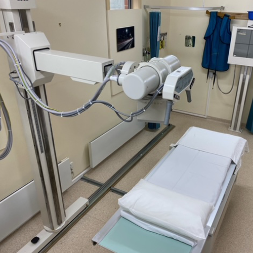
A general radiography (x-ray) can be done on the chest, abdomen, pelvis, skull and limb. It involves exposing a part of the body to a small dose of radiation to produce an image of the internal organs.
When x-rays penetrate the body, they are absorbed in varying amounts by different parts of the anatomy. Ribs and bones, for example, will absorb much of the radiation and, therefore, appear white or light grey on the image. Lung tissue and other internal organs absorb lesser radiation and appear darker on the image. In this manner, a “picture” of the body part is formed
You may be requested to change into an x-ray gown to avoid metallic items, buttons and zippers. You will also be asked to remove jewellery, eyeglasses, and any metal objects that could obscure the image.
Once you are positioned in the required pose with the x-ray plate, you may be asked to take a deep breath and hold it or just to hold your breath and keep still. The radiographer will go to another small room or cubicle and activate the x-ray equipment which will send a beam of x-rays to the positioned area. You need to keep still as any movement will lead to an unsharp picture and an accurate diagnosis cannot be made.
When the x-rays are completed you will be asked to wait until the radiographer examine the images to determine if more are needed.
